 Welcome to the Microscopy Lab! The Microscopy lab offers tools for biological research as well as technical assistance and training. The lab has two major microscopes - a transmission electron microscope (TEM), and a laser-scanning confocal microscope (LSCM) equipped with a multi-photon (MP) laser. The microscopy lab also has both compound and stereomicroscopes for bright field and fluorescence imaging. Ancillary equipment necessary for sample preparation is also available.
Microscopy lab users can request full technical guidance, or once trained, can use the equipment independently. For training and other enquiries, the lab manager is accessible by email at stacy.welker@ufl.edu. To reserve time for your project, scroll down to the listing for the desired equipment and click on the link for the interval you require. If you need to use equipment or reserve an amount of time that is not yet listed, please contact the microscopy lab manager.
Fees
(all charges are per hour)
|
Microscope |
University of Florida |
External Universities |
industry |
|
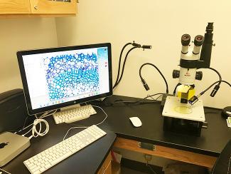
Birghtfield and Dissecting Microscopes |
$15 |
$25 |
$40 |
|
Confocal w/o Multiphoton |
$40 |
$60 |
$80 |
|
Confocal w/ Multiphoton |
$50 |
$70 |
$100 |
|
TEM |
$65 |
$90 |
$130 |
|
Leica Microtome and Cryostat |
$15 |
$25 |
$40 |
|
Ultramicrotomes |
$30 |
$40 |
$50 |
|
Critical Point Drier |
$30 |
$40 |
$50 |
|
Sputter Coater |
$30 |
$40 |
$50 |
Equipment
Transmission Electron Microscope
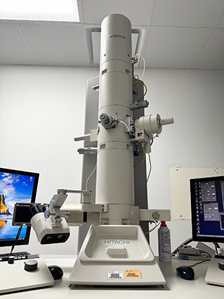
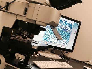
Transmission electron microscopy (TEM) services are performed on our Hitachi H-7600. We upgraded our digital camera to an AMT XR50 5MP CCD camera in the spring of 2017 to increase image quality. The TEM is useful for ultrastructural studies. At the CREC, we predominately use our TEM to study the ultrastructure of various citrus tissue and identify morphological changes as well as pathogens. We also use the TEM in plant-vector interactions. Using the TEM allows us to identify the location of a pathogen in the insect and support transmission hypotheses with morphological studies.
Confocal Laser-Scanning Microscope
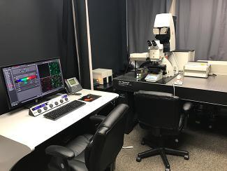
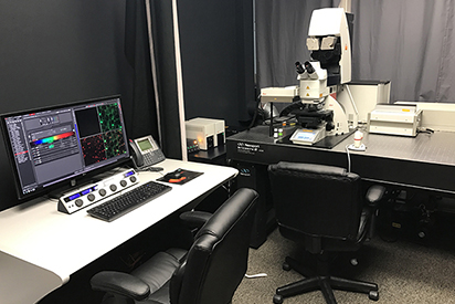
Our Leica SP8 Multi-photon confocal is our newest microscope. This microscope was purchased and installed in Spring of 2016. The confocal uses lasers to illuminate natural auto-fluorescence or detect fluorescing antibodies in a sample. The multi-photon laser can be tuned to excite different molecules at different wavelengths and can often penetrate deeper into tissue than the other lasers used in standard CLSM. Confocal laser-scanning microscopy (CLSM) has advanced our knowledge of cellular interactions. We can use CLSM to study protein-protein interactions, locate expressed genes using In situ hybridization and can even get three-dimensional renderings of organelles, cells, and tissues. The confocal eliminates light above and below the plane of focus to increase the resolution over traditional epi-fluorescence.